biofilm assay protocol
In the protocol described here we will focus on the use of this assay to study biofilm formation by the model organism Pseudomonas aeruginosa. 4- keep without no agitation for 24 or 48 or 72 days until.
Trough base.

. In addition as part of the biofilm inhibition assay. This experiment was then repeated twice to achieve a total of three runs. In this study we have evaluated the impact of methodological approaches in the determination of biofilm formation by four clinical isolates of Escherichia coli in static assays.
The results also demonstrate that cell viability generally correlates with biofilm biomass for-mation and that the CV2 method of biofilm measurement is probably more suitable than the CV1 method. Pipette up and down to assure that the stained biofilm is well solubilized and then transfer 100 μL of each sample to a new 96-well optically clear flat-bottom plate. Our assay kits can reduce measurement variation.
Inoculate biofilm assay plates directly in 100-μl medium per well from the overnight microtiter plate cultures using a sterile 96-prong inoculating manifold. In this method the negatively charged molecules within a biofilm ie. Inoculate 5ml liquid medium with 5µl 1st Overnight culture use disposable test tubes and incubate at proper conditions overnight.
Incubate for 1015 min. 3 Set up four small trays in a series and add 1 to 2 inches of tap water to the last three. Scanning Electron Microscopy SEM.
19162 100case. Staining the Biofilm After incubation dump out cells by turning the plate over and shaking out the liquid. Cover assay plates and incubate at optimal growth temperature for desired amount of time.
4 The peg lid is gently rinsed to removed planktonic bacteria and a serial diluted test solution is dispensed into a new 96-well microplate. General Biofilm Assay Protocol Materials Needed. Early phase biofilms are.
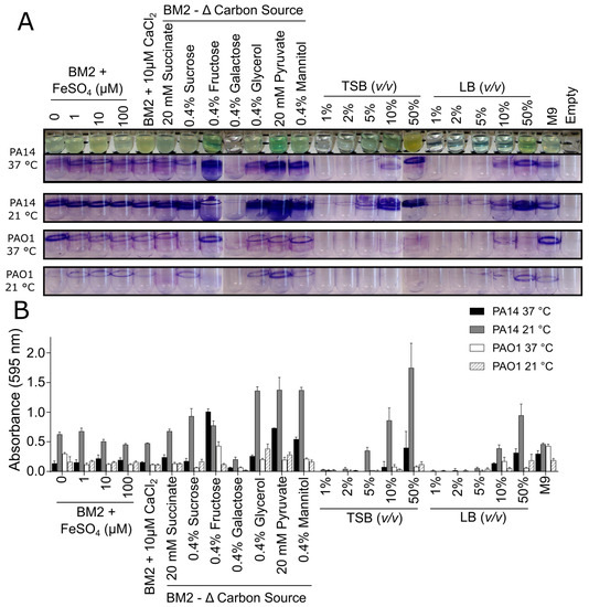
Crystal violet CV assay is the most popular method for biofilm determination adopted by different laboratories so far. For initial assessment A. Polysaccharides of the EPS and.
1 round-bottom 96-well plate 1 flat-bottom 96-well plate Appropriate media Appropriate antibiotic stock 15 mL conical tubes or glass test tubes for growing liquid cultures 01 crystal violet 30 acetic acid Paper towels A large beaker. Aeruginosa biofilm was cultivated on 18 beads according to the standard protocol and processed as described above to determine the number of CFU per bead. Workflow and timeline estimates for the biofilm inhibition gray boxes and eradication white boxes assays for testing the antibiofilm.
A step-by-step summary of the MBEC Assay. Pipette 200 μL of 30 acetic acid solution into each well. Biofilm Assay Protocol for Biofilm assay by Safranin using 96-well plates.
However biofilm layer formed at the liquid-air interphase known as pellicle is extremely sensitive to its washing and staining steps. The assays were performed in microtitre plates with two minimal and two enriched broths with one- or two-steps protocol and using three different mathematical formulas to quantify adherent. The biofilm processing was done as described above.
Pseudomallei biofilms stained with FM1-43 were extracted with various solvents and reagents including methanol and ethanol ranging from 25 to 100 prepared in water 95 isopropanol acetone. In summary the protocol optimized by ImQuest BioSciences provides a high throughput procedure to. In many assays biofilms are quantified by conventional culture plating method to get colony 10 forming unitscount which is an intensive procedure 1 Whereas other assays do use 96 well 11 microtiter plates for biofilm quantification as microtitre plate offers comparatively high 12.
Rapid screening biofilm inhibition assay. To develop an easy to adapt and a robust plate based biofilm quantification assay we optimized a dye extraction protocol post FM1-43 staining. 1-2 Bacteria culture is prepared and dispensed into a 96-well microplate.
In existing methods biofilm formation occurs on the bottom of the well of a microplate which made biofilm easier to peel off because of the processes such as washing. This will solubilize the CV. Products as well as biofilm testing protocols is not available to most industries as there has been minimal biocide testing for microorganisms in the biofilm state in the past.
Incubate the microtiter plate for 4-24 hrs at 37C. In this assay the extent of biofilm formation is measured using the dye crystal violet CV. For quantitative assays we typically use 4-8 replicate wells for each treatment.
1- grow the bacteria 2- guarantee the pure culture 3- next form the biofime add 500 microliters of bacteria into a 24 well microplate. One of the first staining assays used in biofilm analysis was the crystal violet assay. Apply the second overnight culture into plates as your experimental designed pattern and perform the experiment as follow.
33 CV-Stained Biofilm Quantitation 1. MBEC Assay Biofilm Inoculator. This causes measurement variation and this measurement variation was an issue for the quantification of biofilm.
3 The peg lid is placed in the bacteria culture and incubated to generate the biofilm. The protocols can be implemented in most microbiology or immunology research laboratories without the need for specialists.

Biofilm Eradication Testing For Antimicrobial Efficacy

Biofilm Formation Assay Kit Dojindo

A Combination Of Phenotype Microarraytm Technology With The Atp Assay Determines The Nutritional Dependence Of Escherichia Coli Biofilm Biomass Intechopen

Microtiter Dish Biofilm Formation Assay Protocol

Biomolecules Free Full Text Critical Assessment Of Methods To Quantify Biofilm Growth And Evaluate Antibiofilm Activity Of Host Defence Peptides Html

Biofilm Formation Assay Kit Testpiece Dojindo Eu

Quantification Of Biofilm Biomass By Staining Non Toxic Safranin Can Replace The Popular Crystal Violet Sciencedirect

Schematic Representation Of The Steps Involved In The Protocol For Download Scientific Diagram

Biofilm Formation Assay Kit Dojindo

Schematic Crystal Violet Assay On Biofilms In A Microtiter Plate Download Scientific Diagram

Crystal Violet Assay To Assess The Antibiofilm Activity Of Samples Download Scientific Diagram

Biofilm Formation Assay In Pseudomonas Syringae Bio Protocol

Assess The Cell Viability Of Staphylococcus Aureus Biofilms Thermo Fisher Scientific Ca

Biofilm Formation Assay In Pseudomonas Syringae Bio Protocol

Biofilm Formation Assay Kit Dojindo

An Overview Of The High Throughput Protocol For Metal Susceptibility Download Scientific Diagram

Biofilm Formation Assay In Pseudomonas Syringae Bio Protocol
0 Response to "biofilm assay protocol"
Post a Comment